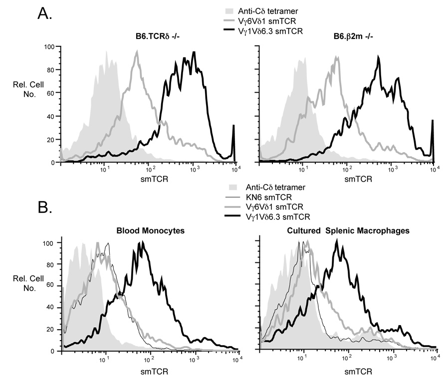
Fig. 6.
A. Freshly isolated peritoneal macrophages from B6.TCRδ−/− vs. B6.β2m−/− mice were stained with either the Vγ1Vδ6.3 or Vγ6Vδ1 smTCR. Macrophages, first identified by staining with F4/80, were then analyzed for staining with each smTCR, as shown. B. Freshly isolated blood monocytes from B6.TCRδ−/− mice were identified by F4/80 staining and any dead cells were excluded by propidium iodide staining; this population was then examined for co-staining with the indicated smTCR. For splenic macrophages, plastic-adherent spleen cells from C57BL/6 mice were cultured overnight before staining. F4/80 positive, propidium iodide negative cells were then analyzed for co-staining with smTCRs as indicated.

