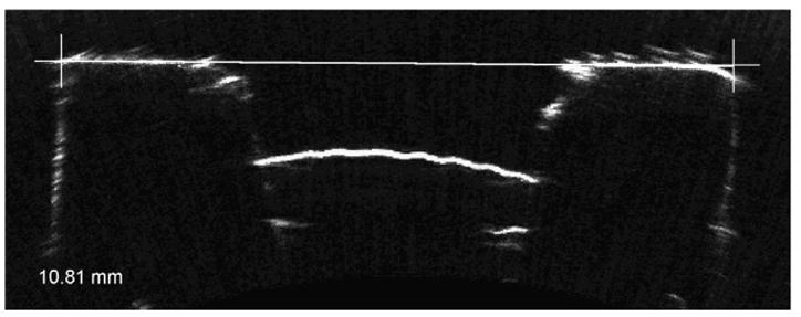
Figure 1.

Horizontal VHF digital US B-scan of the test object (10.80 mm bolt). The shape of the block is seen with the central hole for the screw. The inside of the bolt contains a protruding dome of Blu-Tac. The interfaces appear soft, similar to those seen in US images of the anterior segment. The cross hair shows how the corner was used as a reference point to keep lateral measurements between scans consistent. The white line shows the end points of the measurement that was made by the observer. The distance of the white line is displayed in millimeters in the bottom left-hand corner.
