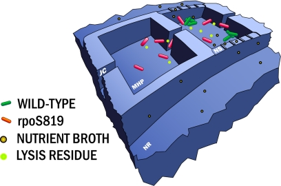
Fig. 1.
Schematic view of 2 adjacent nanofabricated MHPs in a linear array. We constructed fluorescent strains of WT E. coli and GASP mutant (based on the rpoS 819 mutant) to track the populations of competing strains in our landscape. WT bacteria (with green fluorescent proteins) and rpoS 819 mutants (with red fluorescent proteins) are free to move within each MHP and between MHPs through JCs. The NR is coupled to the MHP by NS. The left side habitat patch is closed with all NS closed, and the right side patch is open with full openings. Nutrient and detritus (from bacterial lysis) are denoted by brown and green spheres, respectively, and can diffuse across the NS. Each MHP was 100 μm × 100 μm wide × 8 μm deep; the NS were etched 200 nm; and the feeding channels were 500 μm wide, 200 μm deep, and 1.5 cm long. The net feeder channel volume to MHP volumes ratio was 600:1.

