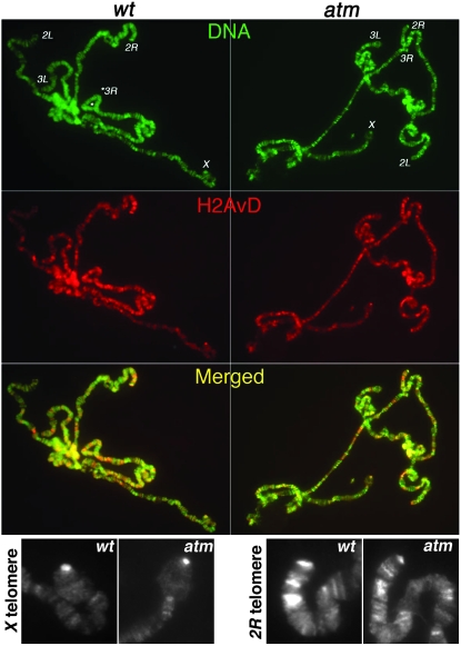
Figure 2.—
H2AvD distribution on polytene chromosomes. DNA signals (DAPI) are in green. Anti-GFP signals are in red. Enrichments of H2AvD-GFP were detected for telomeric regions of chromosome arms X, 2L, and 2R in both wild-type (wt, left) and atm mutant (right panels) cells. (Bottom) Zoomed images of anti-GFP staining at X and 2R telomeres from wild-type and atm mutant cells.

