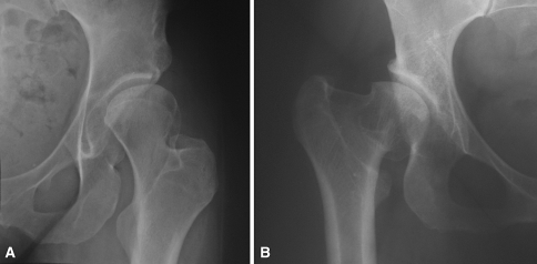
Fig. 2A–B.
Proximal femoral deformities associated with acetabular dysplasia. (A) An anteroposterior radiograph of the left hip in a 31-year-old woman with a history of insidious-onset hip pain demonstrates acetabular dysplasia and associated coxa valga. (B) Anteroposterior right hip radiograph of a 36-year-old female patient with a history of developmental hip dysplasia and previous proximal femoral varus-producing osteotomy shows residual coxa vara.

