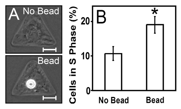
Fig. 5. Proliferation in patterned cells increases upon contact with VE-cadherin-coated beads.
(A) Phase contrast images of solitary spread cells without (top) and with (bottom) a VE-cadherin-coated bead. (B) Graph of BrdU incorporation in spread cells without and with a bead. Error bars are standard error, with (*) p<0.05 versus control by t-test. Scale bars are 10 µm.

