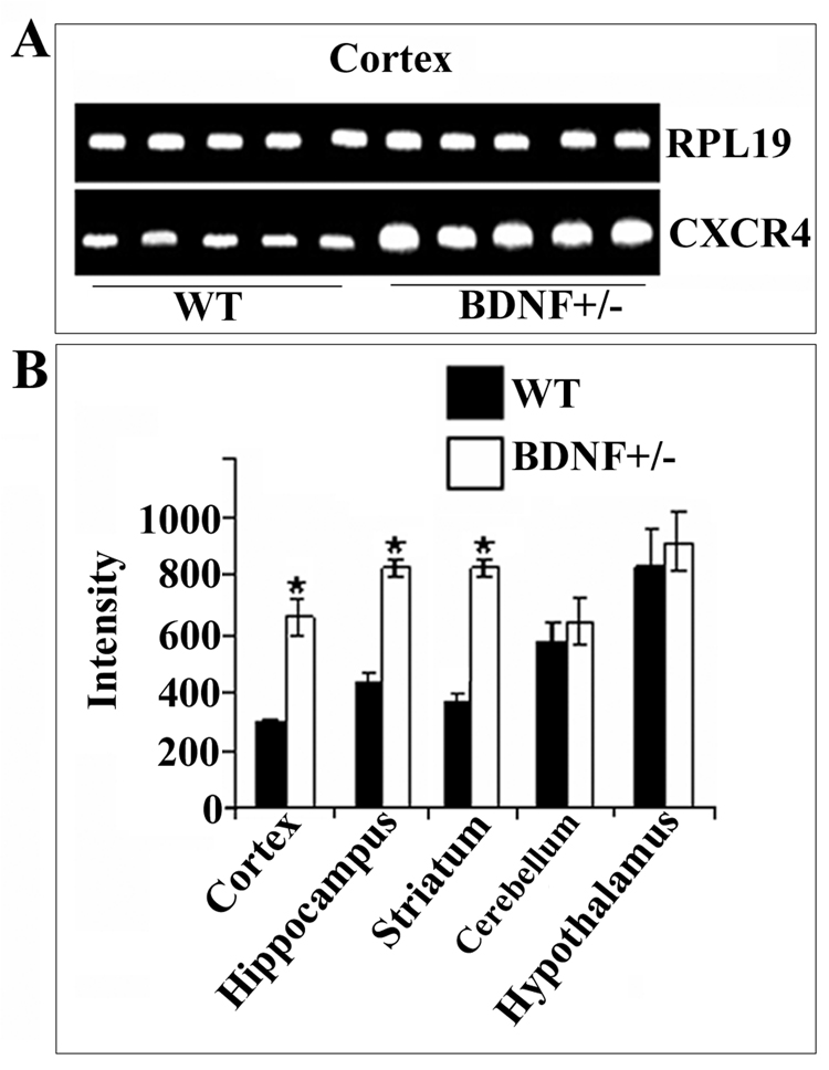
Figure 1. BDNF heterozygous mice exhibit higher levels of CXCR4 mRNA than WT.
RNA was extracted from the indicated brain areas of 6-month-old BDNF+/− mice or WT littermates. RT-PCR was performed using primers designed to amplify mouse CXCR4 (see Experimental Procedures). PCR reaction products were analyzed by agarose gel-electrophoresis (see Experimental Procedures). A. Representative gel showing CXCR4 and RPL19 cDNAs from the cerebral cortex. B. Semi-quantitative analysis of CXCR4 cDNA was carried out by Quantity One 1-D Analysis as described in Experimental Procedures. RPL19 was used as an internal control to normalize gel loading. Data, expressed as intensity of the cDNA band, are the mean ± SEM of five independent samples. *p<0.05 vs control.

