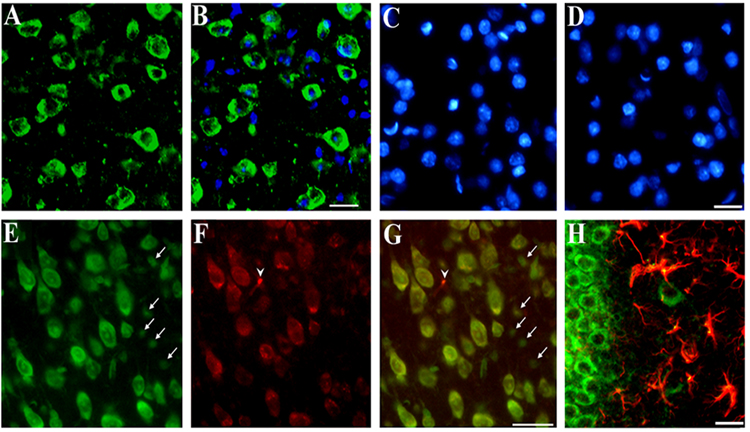
Figure 2. Immunohistochemical analysis of CXCR4.
Serial coronal sections (16 µm) from the frontal cortex or hippocampus were obtained from WT mice. Examples of cortical sections stained with (A) a CXCR4 antibody (green) counterstained with (B) DAPI (blue) to visualize cellular nuclei. Note that some cells are CXCR4 negative. C and D: Examples of sections in which the primary or secondary antibody, respectively, were omitted. Sections were counterstained with DAPI. E and F: Sections stained with Nissl and CXCR4 antibody, respectively (see Experimental Procedures). G: E and F were merged to show colocalization of CXCR4 and neurons (yellow). Note that in E small Nissl positive cells are CXCR4 negative (arrows). In F, one CXCR4 positive cell (arrowhead) is Nissl negative. H: Example of a hippocampal section stained with CXCR4 (green) and GFAP (red) and showing colocalization of CXCR4 and GFAP (yellow). Bars=40 µm.

