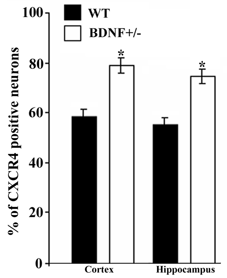
Figure 3. BDNF+/− mice exhibit more CXCR4 immunoreactivity.
Serial coronal sections obtained as described in Fig. 2 and in Experimental Procedures were used to determine the number of CXCR4 positive neurons in the frontal cortex and hippocampus. The number of CXCR4 positive neurons was determined in layer V of the cortex and CA2 region of the hippocampus in an area of 1 mm2 per section using MetaMorph® software. An average of 8000 neurons per animal per area was counted. Data, expressed as % of Nissl positive cells per section, are the mean ± SEM of 10 sections per animal (n=4 each group). *p<0.05 vs WT.

