
Figure 5.
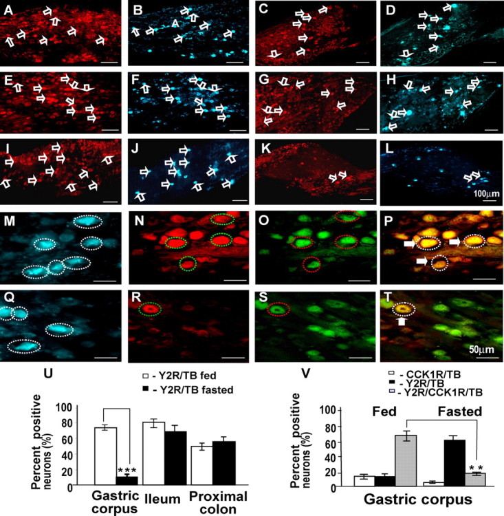
Nodose ganglion neurons projecting to the stomach, ileum, and colon retrogradely labeled with True Blue also express Y2R. A–D, Expression of Y2R in nodose neurons (open arrows) in fed rats projecting to the colon (A) indicated by True Blue fluorescence (B); in fasted rats, there is also Y2R (C) in neurons labeled with True Blue (D) from the colon. E–H, In fed rats, there is Y2R (E) in neurons labeled (open arrows) with True Blue from the ileum (F); in fasted rats, there is also Y2R (G) in neurons labeled with True Blue (H) from the ileum. I–L, In fed rats, there is Y2R in neurons labeled (open arrows) with True Blue from the stomach (I, J), whereas in fasted rats, there is decreased abundance of neurons expressing Y2R (K) and labeled with True Blue from the stomach (L). M–T, At higher power, many neurons (broken circles) retrogradely labeled from stomach in fed animals (M, blue) also contained Y2R (N, red) and CCK1R (O, green) as indicated by the overlay (P; triple-labeled neurons are enclosed in dotted lines and marked by filled arrows). With fasting, neurons projecting to the stomach (Q, blue) only rarely express Y2R (R, red) with CCK1R (S, green) as indicated by the overlay (T, yellow; circled neuron and filled arrow). U–V, Quantification of Y2R-positive vagal afferent neurons projecting to the stomach, ileum, and proximal colon in rats fed ad libitum or fasted. U, Fasting for 48 h decreased the number of neurons projecting to the gastric corpus and expressing Y2R, but expression in neurons projecting to the proximal colon and ileum was unchanged. V, Quantification of Y2R- and CCK1R-immunoreactive neurons projecting to gastric corpus. Compared with rats fed ad libitum in which most neurons serving the stomach expressed Y2R and CCK1R, in fasted rats most did not express Y2R; n = 5; **p < 0.01, ***p < 0.001. Scale bars: 100 μm in A–L and 50 μm in M–T.