Figure 5.
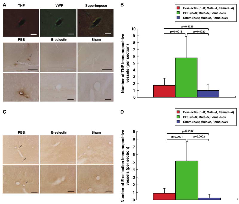
Suppression of vessel activation and local TNF production by mucosal tolerance to E-selectin. (A) Photomicrographs of double-labeled fluorescent immunohistochemistry with anti-TNF and anti-von Willebrand factor antibodies in corpus callosum blood vessels. Immunoreactive TNF was expressed in and around endothelial cells. In the E-selectin-tolerized and sham-operated animals, TNF-immunopositive vessels were markedly less prominent and less frequent than in PBS-treated animals. Scale bars, 50 μm (top); 300 μm (middle), and 50 μm (bottom). (B) Histograms of the numerical density of TNF-immunopositive vessels in the corpus callosum. In the E-selectin-treated and sham-operated animals, the number of TNF-immunopositive vessels was significantly reduced, as compared with the PBS-treated animals. (C) Photomicrographs of the immunohistochemical staining for E-selectin in the corpus callosum. In the E-selectin-treated and sham-operated animals, E-selectin-immunopositive vessels were less prominent and less frequent compared with the PBS-treated animals. Scale bars, 100 μm (top) and 50 μm (bottom). (D) Histograms of the numerical density of E-selectin-immunopositive vessels in the corpus callosum. In the E-selectin- and sham-operated animals, the number of E-selectin-immunopositive vessels was significantly reduced compared with the PBS-treated animals.

