
Figure 5. Phosphorylation of p38 occurs in distinct subcellular compartments following BCR and TLR9 stimulation.
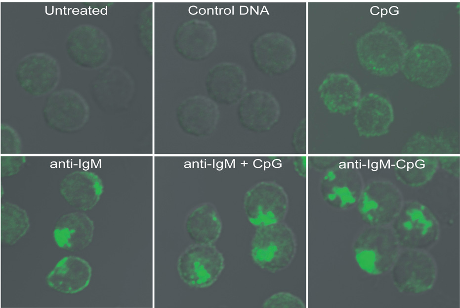
Mouse splenic B cells were either untreated or incubated with: 3µM control GpC DNA; 3µM stimulatory CpG DNA alone; 10µg/ml anti-IgM alone; both 10µg/ml anti-IgM and 3µM CpG DNA; or 10µg/ml anti-IgM-CpG DNA conjugate for 60 min. In each case the cells were fixed, permeabilized and stained for p-p38 using rabbit antibodies specific for p-p38, followed by Alexa 488-labeled goat antibodies specific for rabbit Ig. Shown are the DIC images merged with confocal images of Alexa 488. Approximately 250 cells were analyzed for each condition in 6 independent experiments and representative images are provided.