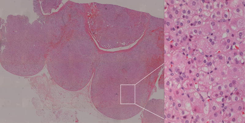
Figure 4. Pathology: MAH.
Digitally processed photomicrographs of adrenal glands with hematoxylin and eosin stain. Left panel: MAH with multiple nodules and hyperplasia throughout the section. Entire tissue section imaged with Mosaix© software, 10X. Right panel: Non-pigmented nodule. 40X magnification of area surrounded by box in left panel.

