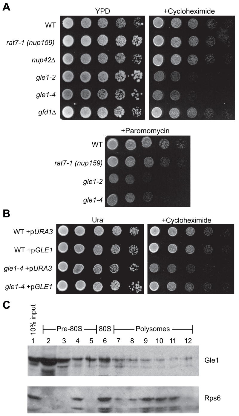
Figure 1. A role for Gle1 in translation.
(A) Cultures of gle1-2, gle1-4, rat7-1 (nup159), nup42Δ, gfd1Δ, and wild-type (WT) strains were spotted in five-fold serial dilutions on YPD alone or YPD containing 0.1 μg/ml cycloheximide or 0.4 mg/ml paromomycin, then incubated 2 days at 23°C.
(B) Cultures of gle1-4 and wild-type strains transformed with empty URA3/CEN plasmid (pURA3) or a GLE1/URA3/CEN (pGLE1) plasmid were spotted as in (A) on Ura− selective media with or without cycloheximide.
(C) Lysates of wild-type strains grown at 23°C were subjected to sucrose density fractionation and immunoblotted with α-Gle1 and α-Rps6 (ribosomal protein control). Ribosome distribution was determined by OD254.

