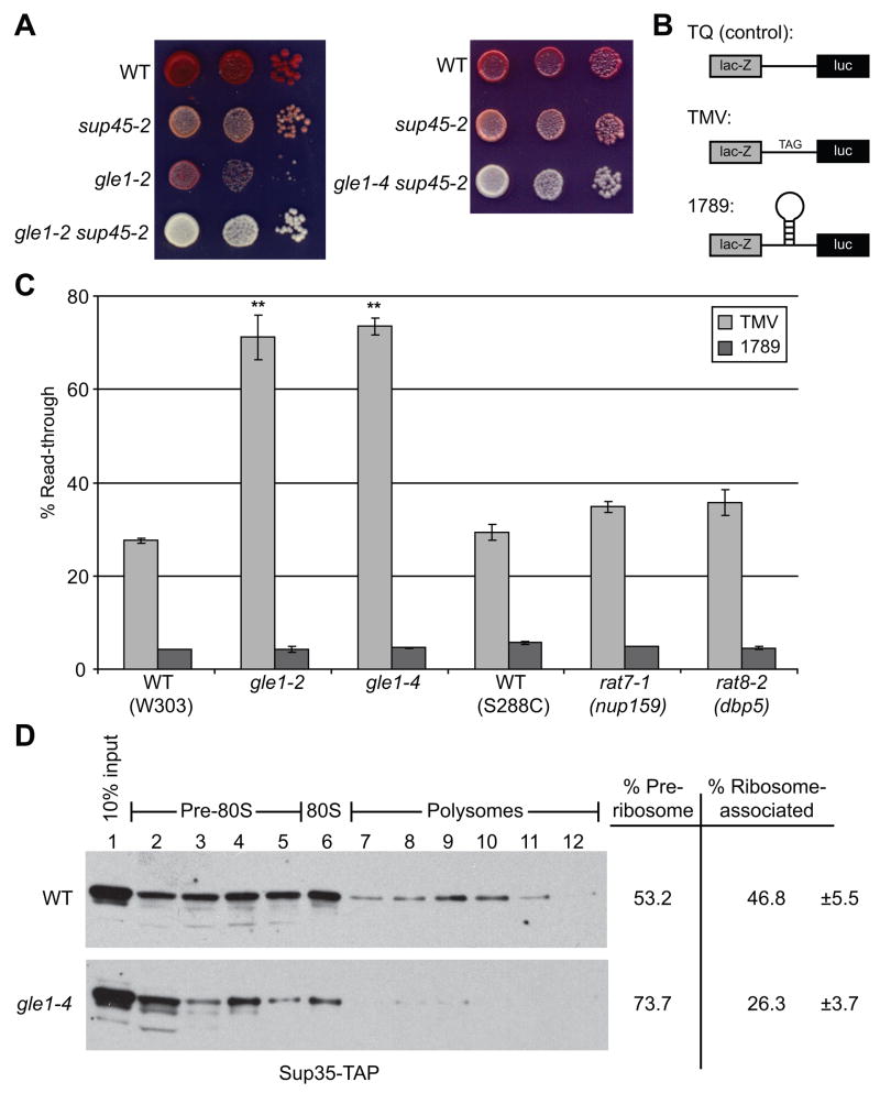
Figure 3. Gle1 is required for efficient translation termination.
(A) Cultures of gle1-2, sup45-2, gle1-2 sup45, gle1-4 sup45-2 and wild-type controls (WT) were serially diluted and spotted on 0.25 YPD. Plates were incubated 4 days at 23°C and colony color assessed.
(B) The tandem β-galactosidase/luciferase reporter constructs are shown: TQ (control) lacks a stop codon in the linker, TMV has a stop codon inserted in frame into the linker region, and 1789 has a stem-loop from HIV-1 inserted into the linker.
(C) gle1-2, gle1-4, rat8-2 (dbp5), rat7-1 (nup159), and wild-type control (WT) strains were transformed with the tandem reporter constructs and luciferase and β-galactosidase assays were performed. Activities were normalized to cell number, and ratios of luciferase to β-galactosidase activity determined. Read-through activity is expressed as the percentage of the TMV or 1789 reporter compared to TQ control for each strain, n = 3–5 independent experiments. Error bars = 1 SEM above and below the mean. ** p<0.01
(D) Lysates of SUP35-TAP and gle1-4 SUP35-TAP strains grown at 23°C were subjected to sucrose density fractionation and immunoblotted with α-mouse IgG to detect Sup35-TAP. Ribosome distribution was determined by OD254, and blots were analyzed by densitometry to assign ribosome association percentages (n = 4). Error = 1 SEM above and below the mean. p<0.05

