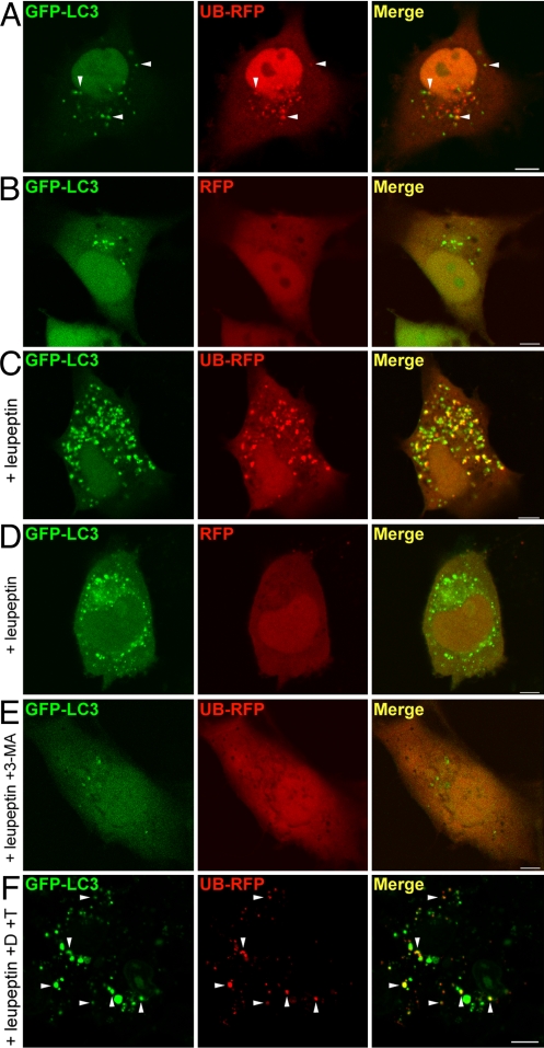
Fig. 1.
Ubiquitin targets cytosolic RFP to autophagosomes for degradation. (A and B) COS-7 cells transiently cotransfected with GFP-LC3 and UB-RFP (A), or GFP-LC3 and RFP (B). Cells were imaged 24 h after transfection. Arrowheads in (A) indicate obvious examples of colocalized GFP-LC3 and UB-RFP. (C and D) COS-7 cells transiently cotransfected with either GFP-LC3 and UB-RFP (C) or GFP-LC3 and RFP (D), and treated with 0.25 mM leupeptin for 20 h before imaging. (E) COS-7 cells transiently coexpressing GFP-LC3 and UB-RFP, and treated with 0.25 mM leupeptin and 10 mM 3-MA for 20 h before imaging. (F) Fluorescence Protease Protection assay of COS-7 cells coexpressing GFP-LC3 and UB-RFP. 24 h after cotransfection and leupeptin treatment, cells were washed and then treated with 0.6% [vol/vol] digitonin (D) for 10 min and than with 0.005% [wt/vol] trypsin (T), followed by imaging. Arrowheads indicate obvious examples of colocalized GFP-LC3 and UB-RFP. (Scale bars, 10 μm.)

