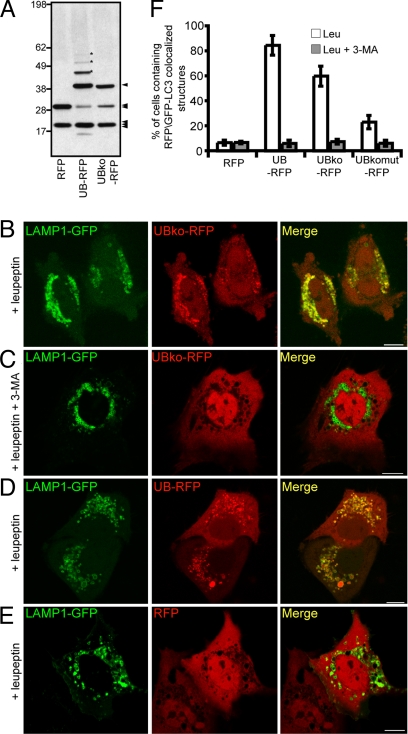
Fig. 2.
Monomeric ubiquitin is sufficient to target RFP for degradation by autophagy. (A) Immunoblot of cell lysate from COS-7 cells expressing RFP, UB-RFP, or UBko-RFP. 25 μg of total protein was resolved by SDS/PAGE and immunoblot using rabbit anti-RFP antibodies and donkey anti-rabbit IgG conjugated to horseradish peroxidase. Asterisks indicate the position of three higher molecular weight species of polyubiquitinated UBko-RFP. The single arrowhead indicates the position of mono-ubiquitinated RFP, whereas the double and triple arrowheads indicate the position of RFP alone and a cross reacting band, respectively. Molecular masses (in kDa) are indicated on the left side of the blot. (B–E) COS-7 cells cotransfected with LAMP1-GFP and either UB-RFP (B and C), UB-RFP (D), or RFP (E). Leupeptin (0.25 mM) was added 20 h before imaging. Cells shown in (C) were treated also with 10 mM 3-MA. (Scale bars, 10 μm.) (F) Quantification of the percentage of cells with five or more punctate RFP signals that colocalized with GFP-LC3 in cells coexpressing GFP-LC3 and various RFP constructs as indicated. Cells were also incubated with either leupeptin alone (white bar), or with leupeptin and 3-MA (dark gray bars). Shown are the averages ± standard deviations from three independent experiments with each experiment including at least 50 cells scored.

