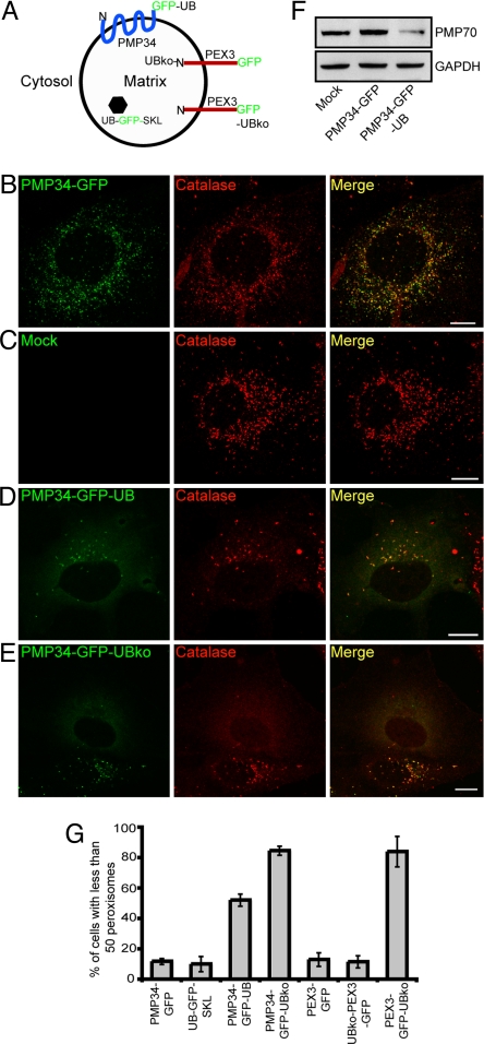
Fig. 4.
Ubiquitination of a PMP results in a decrease in the number of peroxisomes within a cell. (A) Schematic illustration of the predicted topological orientation of PMP34-GFP-UBko, UB-GFP-SKL, UBko-PEX3-GFP, and PEX3-GFP-UBko. (B–E) COS-7 cells transiently expressing either PMP34-GFP (A), empty vector (mock) (B), PMP34-GFP-UB (C), or PMP34-GFP-UBko (D) were fixed and stained with anti-catalase and Alexa 543-goat anti-rabbit antibodies 48 h after transfection. Note that two PMP34-GFP-UBko-transformed cells can be seen in (D), both of which display a reduced number of peroxisomes relative to cells transformed with the empty vector (B) or PMP34-GFP (A). (F) Immunoblot of cell lysate from COS-7 cells expressing PMP34-GFP, PMP34-GFP-UB or mock treated. Cells were lysed, and 25 μg of total protein was subjected to SDS/PAGE and then immunoblotted with antibodies against PMP70 or the cytosolic protein glyceraldehydes phosphate dehydrogenase (GAPDH) serving as a protein loading control. (G) Percentage of total number of transfected cells with less than 50 peroxisomes per cell expressing various proteins as indicated 48 h after transfection. Shown are the averages ± standard deviations from three independent samples with each experiment including at least 100 cells scored.

