Fig. 6.
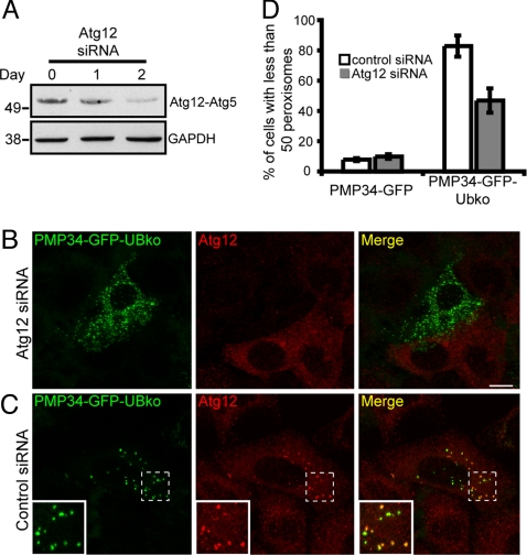
Silencing of Atg12 expression prevents ubiquitin-mediated peroxisome degradation. (A) Immunoblot of cell lysate from HeLa cells before Atg12 siRNA treatment (day 0), or 1 and 2 days after Atg12 siRNA treatment as indicated. Cells were lysed and 25 μg of total protein was subjected to SDS/PAGE and then immunoblotted with antibodies against either Atg12 or GAPDH serving as a loading control. Depletion in Atg12 expression was indicated by the decrease in the Atg12-Atg5 protein conjugate. (B–C) HeLa cells were transfected with either siRNA pool directed against Atg12 (B), or a nontargeting control siRNA (C). Twenty-four hours after the initial transfection, cell were transfected again with the appropriate siRNA along with a plasmids encoding PMP34-GFP-UBko. Forty-eight hours after the second transfection cells were fixed and stained with anti-Atg12 and Alexa 543-goat anti-rabbit antibodies. (D) Percentage of the total number of cells with less than 50 peroxisomes per cell expressing either PMP34-GFP or PMP34-GFP-UBko and treated with either control siRNA or Atg12 siRNA. Shown are the averages ± standard deviations from three independent samples with each experiment including at least 100 cells scored. (Scale bars, 10 μm.)

