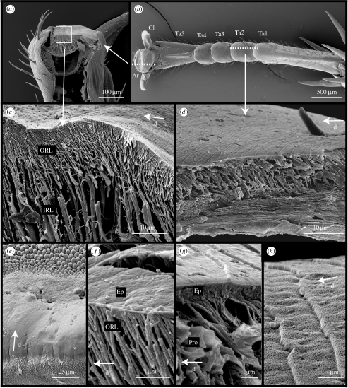
Figure 1.
Morphology of arolium and euplantulae in N. cinerea. (a,b) Arrangement of attachment structures on the tarsus. (c) Freeze fracture of the arolium contact zone showing branched cuticle fibres. (d) Cuticle structure of the euplantulae. (e) Surface morphology of the arolium. (f) Epicuticle of the arolium and fine rods of the procuticle. (g) Epicuticle of the euplantulae and procuticular rods. (h) Surface profile of the euplantulae. Ar, arolium; Cl, claws; PrT, pretarsus; Ta1-5, tarsal segments 1–5; ORL, outer rod layer; IRL, inner rod layer; Ep, epicuticle; Pro, procuticle; d, distal direction.

