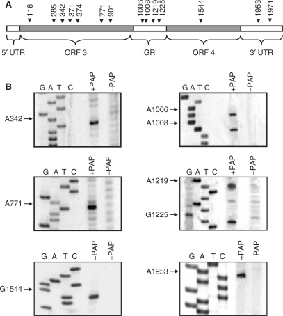
Figure 2.
In vitro depurination of BMV RNA3 by PAP. (A) Schematic of BMV RNA3 indicating the sites of depurination by their nucleotide number. The 5′ untranslated region (5′ UTR; 1–91 nt), the open reading frame of RNA3 (ORF3; 92–1003 nt), the intergenic region (IGR; 1004–1246 nt), the open reading frame for RNA4 (ORF4; 1247–1813 nt) and the 3′ untranslated region (3′ UTR; 1814–2113 nt) are also indicated. (B) Representative depurination sites determined by primer extension. In vitro transcript of RNA3, either PAP-treated or untreated, was extended with reverse transcriptase using cDNA primers distributed over the length of the viral RNA. Radiolabeled cDNA products were separated in a 7M urea/6% acrylamide gel and visualized by autoradiography. The same primers were used to identify the depurination sites by deoxynucleotide sequencing of BMV DNA3.

