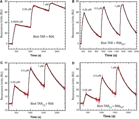
Figure 4.
Kinetic analysis of TAR-aptamer complex formation by surface plasmon resonance. A total of 80–90 RU of biotinylated TAR and TARC5 were immobilized onto streptavidin-coated SA sensorchips (BIAcore™). Hairpin aptamers, prepared in the running buffer, were injected across the sensor surface at a 20 µl/min flow rate, at 23°C. The sensorgrams were fitted as described in Materials and Methods section assuming a pseudo-first kinetic model of the aptamer binding to the immobilized TAR hairpins. The red curves represent the recorded data and the black one the fit of the sensorgrams to a kinetic titration dataset of three analyte injections. (A) Injection of the unmodified R06 aptamer (0.0625 µM, 0.25 µM and 1 µM) across the TAR-coated flowcell. (B) Injection of R06G5* (0.25 µM, 0.5 µM and 1 µM) across the TAR-coated flowcell. (C) Injection of the unmodified aptamer (0.25 µM, 0.5 µM and 1 µM) across the TARC5-coated flowcell. (D) Injection of R06G5* (0.25 µM, 0.5 µM and 1 µM) across the TARC5-coated flowcell.

