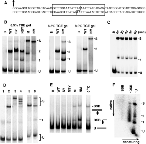
Figure 6.
Binding and synapsis of site I by resolvase variants. (A) The site I-containing double-stranded oligonucleotide used in the binding and synapsis experiments. The site I sequence is boxed. The staggered line indicates the bonds cleaved by resolvase. The top strand was 5′-end labelled with 32P (asterisk). (B) Binding and synapsis by wild-type (WT) resolvase and activated mutants, assayed by non-denaturing polyacrylamide gel electrophoresis. Lanes marked B are controls (incubated without resolvase). Bands corresponding to unbound site I DNA, complexes containing 1 or 2 resolvase subunits and site I synapse are indicated by U, 1, 2 and S, respectively [band assignment based on (14,34)]. The SY resolvase used here has a C-terminal hexahistidine tag whereas all the other resolvases do not, which might account for the slightly lower mobilities of the SY complexes. Left panel; 6.5% polyacrylamide gel with TBE buffer. Centre panel; 6.5% gel with TGE buffer. Right panel; 8% gel with TGE buffer. (C) Time course of site I binding/synapsis by NM resolvase. Resolvase was added to samples at 0°C, and aliquots were withdrawn from the mixture at the stated times, then loaded on a running 6.5% TBE gel. (D) Assembly of the site I synapse with different sized resolvases. NM–GFP is NM resolvase with a 23 kDa GFP domain fused to the C-terminus. Binding/synapsis mixtures were separated on a 6.5% TBE gel. Lane 1, no resolvase. Lane 2, +NM resolvase. Lane 3, +NM–GFP resolvase. Lane 4, +a 1:1 mixture of NM and NM–GFP. Lane 5, NM resolvase (400 nM) was added to site I, at 22°C. After 5 min, NM–GFP (400 nM) was added, and the sample was loaded on the gel after a further 10 min. Lane 6, NM and NM–GFP resolvases were added separately to site I, and the two samples were mixed after 5 min at 22°C. The mixture was loaded on the gel after a further 10 min. (E) Cleavage of site I by activated resolvase mutants. Reactions were set up as in (B), and after 30 min at 37°C the resolvase was denatured with 0.1% SDS. Electrophoresis was on a 6.5% polyacrylamide gel in TBE buffer containing 0.1% SDS. The positions of bands containing unbound DNA (U), and DNA with a SSB DSB are indicated. A resolvase subunit is covalently attached to the SSB and DSB species. In the lane on the right, NM resolvase was added to site I at 0°C and 0.1% SDS was added after 5 min. (F) 2D analysis of site I synapse. A sample containing NM resolvase and site I was separated on a 6.5% polyacrylamide–TBE gel, then soaked in SDS-containing buffer to denature resolvase and run in a second dimension in the presence of SDS. Annotation of the gel is as in parts B and E.

