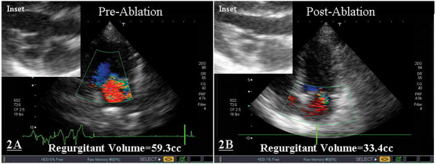
Figure 2. Pre- and Post-Ablation Morphology and Structure of Myxomatous Mitral Valve.
Transthoracic echocardiograms performed before (2A) and after RFA (2B) demonstrated a reduction in the degree of MVP, MR, and chordal length (see insets). This animal underwent 3 applications of RFA at an average power of 12.2±1.4W for an average of 50±10sec each.

