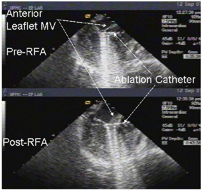
Figure 3. ICE-Guided Percutaneous Ablation of Mitral Valve Anterior Leaflet.

To guide mitral valve ablation, ICE catheter was placed in the right atrium and positioned to give a modified three chamber view. The left atrium is a 12 o’clock, the left ventricle is directly inferior to the left atrium, and the ablation catheter is advanced retrograde across the aortic valve and positioned adjacent to the anterior leaflet of the mitral valve. The top image depicts the mitral valve prior to RFA and the bottom image depicts the mitral valve following RFA. One notes the obvious thickening found post-RFA.
