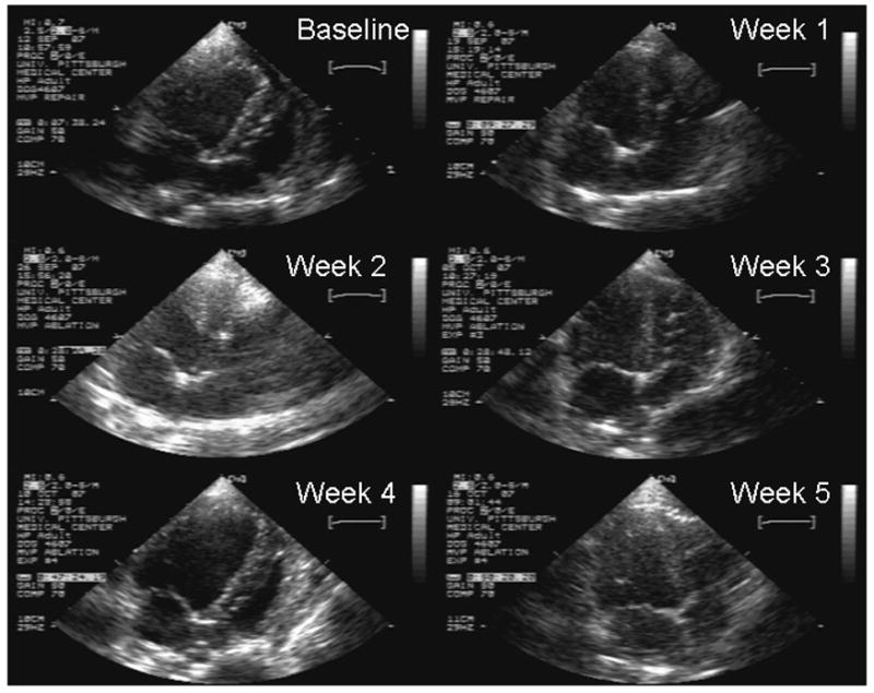
Figure 4. Chronic Echocardiographic Changes after Percutaneous Anterior Mitral Leaflet Ablation.

The transthoracic apical 4 chamber echocardiogram still frames were taken at baseline and weekly thereafter for 5 weeks. The baseline echo demonstrates a typical baseline mitral valve morphology. The subsequent echocardiograms depict the typical changes found after RFA. One can see thickening of the entire anterior leaflet, pronounced at the base and tip of the leaflet that persists throughout the entire follow-up.
