Fig. 2.

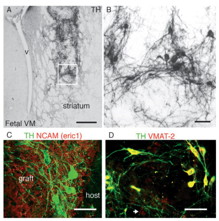
(A and B) Tyrosine hydroxylase (TH) immunoreactivity in a representative graft from primate fetal ventral midbrain at 16 weeks post-transplantation into the striatum of a hemi-parkinsonian 6-OHDA-lesioned rat. The boxed area is magnified in (B). (C) Primate cells in the graft were identified by the primate specific marker neural cell adhesion molecule (NCAM; eric 1, red), TH + (green) neurons within the graft were located in clusters, close to the host–graft interface, and (D) co-expressed the vesicular monoamine transporter (VMAT-2, red). VMAT-2 +/TH – neurons were also present in these grafts (arrow). V, ventricle; Scale bar: 500 μm (A); 50 μm (B–D).
