
Fig. 3.
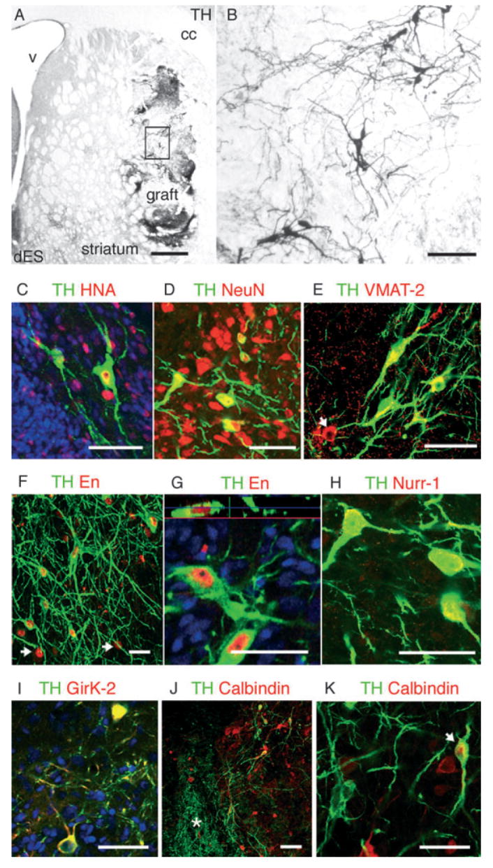
(A and B) Tyrosine hydroxylase (TH) immunoreactivity in a representative graft derived from differentiated primate ES cells (dES) showing TH + neurons within the graft (boxed area is magnified in B) and areas of dense TH neuritic arborization. (C) ES-derived TH + (green in C–K) neurons in the grafts expressed the primate-and human-specific nuclear antigen (HNA, red), (D) neuronal nuclear antigen (NeuN, red) and (E) vesicular monoamine transporter (VMAT-2, red). Like in the fetal grafts (Fig. 2D), VMAT-2 +/TH – cells were also found (arrow). (F) TH + neurons in dES grafts expressed the homeobox transcription factor Engrailed (En, red, 93 ± 3%) and ~45% of En + cells expressed TH (arrows in F point to En +/TH – cells) (G) Nuclear localization of En (red) in a TH + neuron is shown in an orthogonal reconstruction of serial confocal optical sections. (H) Nurr1 (red) was also expressed by TH + neurons. (I) Some TH + neurons in the grafts co-expressed the G-coupled inward rectifier K+ channel 2 (Girk-2, red), which is preferentially expressed by midbrain dopamine neurons in the substantia pars compacta. (J and K) Some TH + neurons co-expressed calbindin (red) (J, arrow in K), which is highly expressed in the A10 dopamine subpopulations. Calbindin +/TH – neurons were also found in the grafts, but not in the areas of dense TH innervation (asterisk in J, see Fig. 4). cc, corpus callosum; V, ventricle. Scale bars: 500 μm (A); 50 μm (B–K).