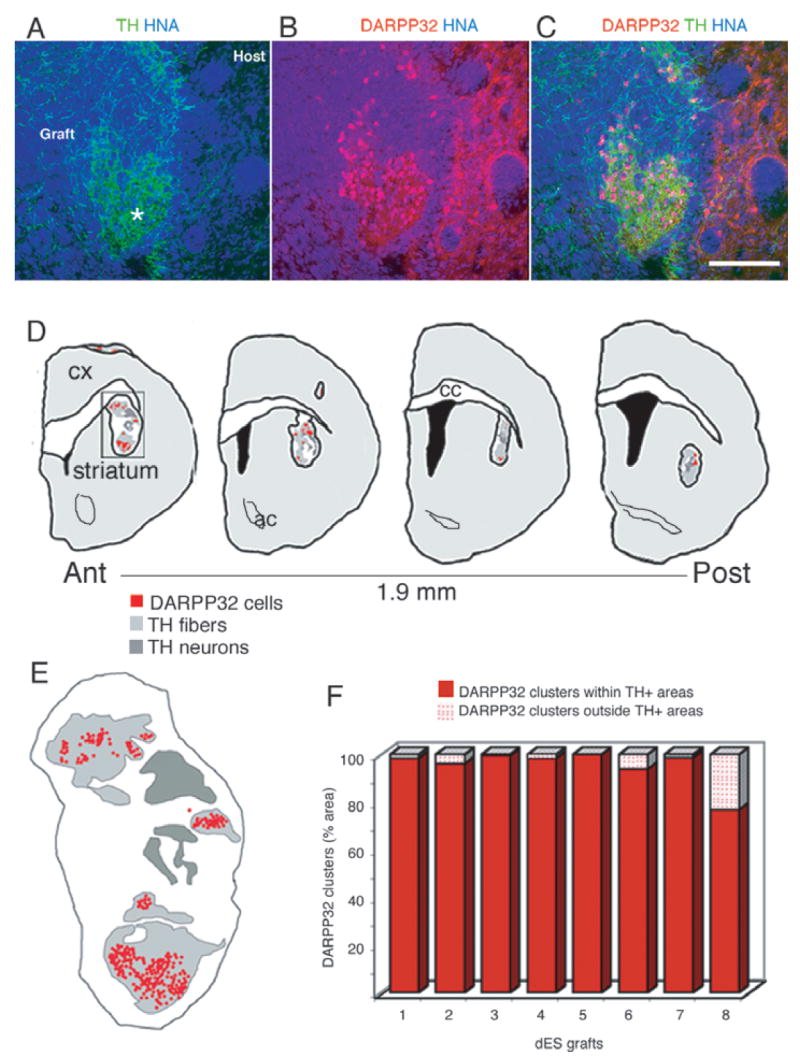
Fig. 7.

(A) Patches of dense tyrosine hydroxylase (TH) + (green) fibers within the graft [human nuclear antigen (HNA) +, blue] in a representative animal transplanted with dES. (B) Primate dopamine and cAMP-regulated phosphoprotein (DARPP)-32 + neurons (red, overlap with HNA appears purple) were found in clusters that (C) coincided with the areas of TH + innervation. (D and E) In order to quantify this distribution, DARPP-32 + neurons and TH + areas were mapped on serial sections (480 μm apart) spanning the whole graft in animals grafted with dES (n = 8): maps of a representative graft showing the distribution of DARPP-32 + neuronal clusters with respect to TH innervation. (E) Detail of the map boxed in (D): each red dot represents one primate DARPP-32 + neuron; the regions densely innervated by TH + fibers are outlined in light gray (see A and Fig. 3B, asterisks), and the regions in dark gray delineate the areas where the TH + cell bodies are (for morphological delineation of these areas, see Fig. 3A and B). (F) Quantification of DARPP-32 + neurons distribution was done on each map, and the percentage of DARPP-32 + neurons present inside and outside TH-innervated areas is shown for each animal. Almost all (95 ± 2.7%) DARPP-32 + neurons were located within TH-innervated areas, supporting a strong attractive effect of DARPP-32 + neurons and TH + fibers. Scale bar: 150 μm.
