
Figure 1. Structure of the VP1 apoenzyme.
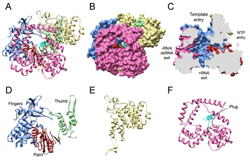
Ribbon diagram (A) and surface rendering (B) of the complete VP1 polypeptide chain. The N-terminal domain is in yellow, the C-terminal (bracelet) domain in pink, and the C-terminal plug in cyan. The conventionally designated subdomains of the polymerase domain are in light blue (fingers), red (palm), and green (thumb). (C) Sagittal cutaway of the image in (B), after rotation to the left by 90°, showing the four tunnels extending into the central cavity. (D-F) Ribbon diagrams of the VP1 domains: polymerase, N-terminal, and C-terminal domains.