
Figure 5. Comparison of the VP1/3′CS+ and the λ3 initiation complexes.
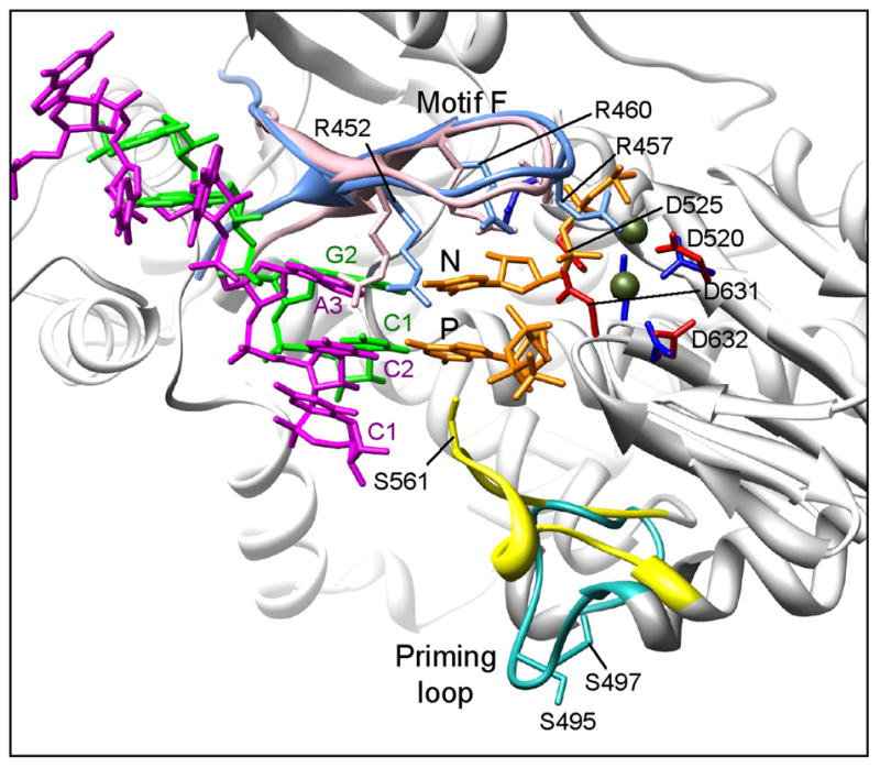
Elements of the rotavirus VP1/3′CS+ complex are superimposed on a ribbon representation of the reovirus λ3 initiation complex (light gray). The priming (P) and incoming (N) nucleotide positions are indicated, and Mg2+ ions are shown in olive. The template RNAs of rotavirus (UGUGACC, magenta) and reovirus (UAGC, green) and the incoming nucleotides (orange) are shown. The priming loop (λ3, yellow; VP1, turquoise) and motif F (λ3, pink; VP1, light blue) are colored, and the location of several key residues, including the catalytic aspartates (λ3, dark blue; VP1, red) and motif F arginines are indicated.