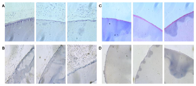
Figure 10.

(A) Hematoxylin and eosin staining of subcutaneous LysB10 implants demonstrates the presence of a mild foreign body reaction along the periphery. (B) F4/80 staining of subcutaneous LysB10 implants demonstrate the presence of macrophages along the periphery of the fibrous capsule. (C) H&E staining of peritoneal LysB10 implants demonstrates the presence of a mild foreign body reaction along the periphery. (D) F4/80 staining of peritoneal LysB10 implants demonstrates the presence of macrophages along the periphery. Images are oriented so that the LysB10 gel is located in the bottom right corner, 20x magnification.
