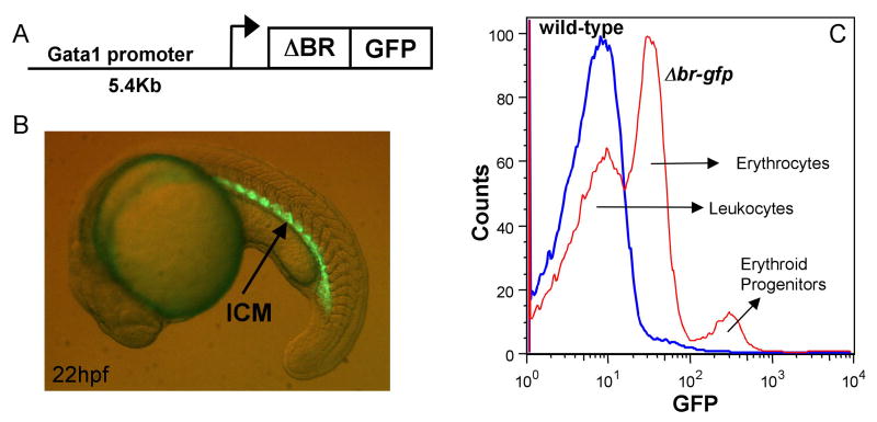
Fig. 6. A mutant receptor isoform is expressed in progenitors of a gata1:Δbr-gfp transgenic line.
(A) The diagram represents the construct used to generate the gata1:Δbr-gfp transgenic line. A 5.4 kb promoter sequence was cloned upstream of a fusion construct encoding a truncated BMP receptor with a C-terminal GFP tag. (B) A representative example of a gata1:Δbr-gfp embryo at 22 hpf viewed under fluorescence. The embryo shows GFP expression (arrow) in the intermediate cell mass (ICM), which recapitulates the endogenous gata1 expression pattern. GFP expression is significantly down-regulated as the cells differentiate (not shown). (C) WKM from homozygous adult gata1:Δbr-gfp fish was analyzed by FACS along with WKM from wild-type (non-transgenic) fish. Samples show three distinct patterns for GFP expression. An overlay histogram of GFP expression is shown and the corresponding cell types listed, wild-type (blue), and gata1:Δbr-gfp (red). Wild-type WKM cells have no GFP expression. The gata1:Δbr-gfp WKM shows a different profile of GFP expression in mature erythrocytes compared to gata1:gfp adults, because the fusion protein is down-regulated as erythroid cells differentiate. GFP is expressed at a low level in the progenitor cells of gata1:Δbr-gfp, while very low levels of GFP are expressed in the mature erythrocytes. These cell types were confirmed by sorting and cytospins (data not shown). Although not shown here (see Fig. 1B) WKM from gata1:gfp adults shows high GFP expression in mature erythrocytes and low GFP expression in the progenitors.

