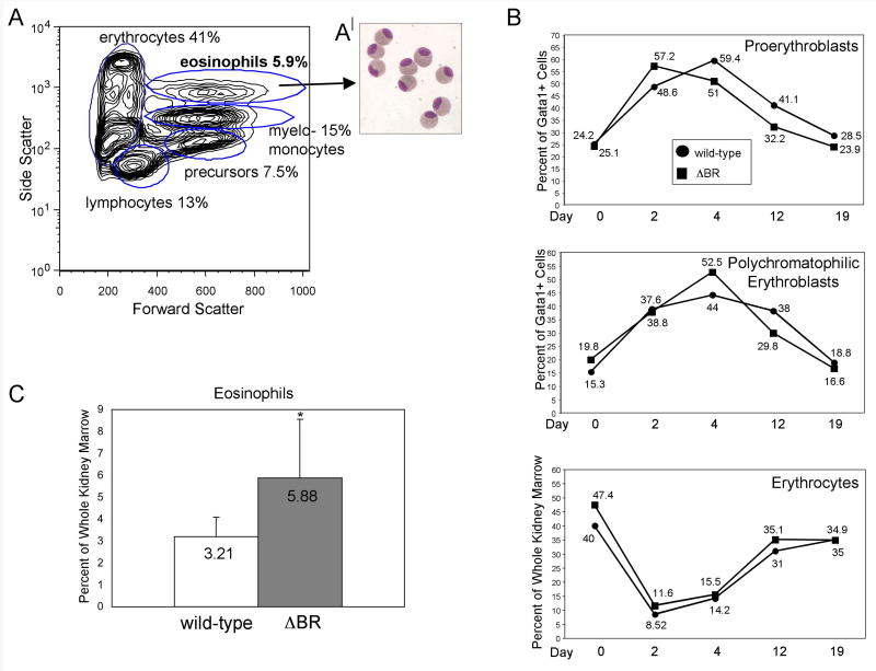
Fig. 7. The gata1:Δbr-gfp adults have a normal response to stress anemia, but increased eosinophil production in WKM.
(A) A contour plot from FSC vs. SSC analysis of gata1:Δbr-gfp adult WKM is shown. While the cell numbers of the four major sub-populations are relatively normal, a fifth side scatter high (very granular) population of cells can be discriminated in these fish, representing eosinophils. (B) Cohorts of wild-type (gata1:gfp) and gata1:Δbr-gfp adults were treated with phenylhydrazine at day 0. On days 2, 4, 12 and 19 adults were sacrificed and WKM prepared and analyzed by FACS. Dead cells were gated out using DAPI staining, and the live cells were analyzed by forward scatter, side scatter and GFP. The percent of each sub-population of erythroid progenitors was determined by gating on either the lymphoid or precursor fraction and then determining the number of GFP(low) positive in that fraction. No significant changes were seen when comparing wild-type and gata1:Δbr-gfp fish. For each time point n=4. As indicated in the insert: ●=wild-type ■=gata1:Δbr-gfp. (C) Comparison of WKM sorts with wild-type (gata1:gfp) shows a relative increase in the number of eosinophils in WKM of gata1:Δbr-gfp adults; for each n=16, P<0.007. (A′) This population of cells was collected, centrifuged onto slides and stained with Giemsa and May-Grünwald to confirm their identification as eosinophils.

