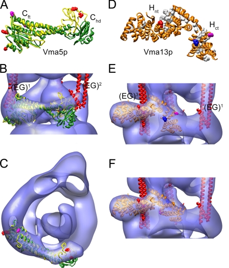
FIGURE 6.
Fitting of subunit C and H crystal structures into the yeast V-ATPase three-dimensional model. A, overlay of the high (green) and low (yellow) conformers of subunit C (PDB 1u7l; Ref. 42). As can be seen, the foot domains (Cft) match well, whereas the position of the head domains (Chd) varies, indicating some flexibility of the two domains. Coordinates of the low-resolution conformer were kindly provided by Dr. Nathan Nelson. The sites from where cross-linking to EG has been observed (34) are in red spacefill. B and C, fitting of both subunit C conformers into the V-ATPase three-dimensional model reveals a better fit for the low-resolution conformer. D, crystal structure of subunit H (PDB 1ho8; Ref. 43) showing an N-terminal domain (Hnt) and a C-terminal domain (Hct). Conserved residues are in white spacefill. Residues from where cross-links to subunits E, B, and F have been observed (54) are in red, blue, and magenta spacefill, respectively. E, fitting of the crystal structure of subunit H into the V-ATPase three-dimensional model. In this orientation, the sites cross-linking to B and F are on the outside. The Hct domain was therefore fitted separately (see panel F) in accordance with the independent behavior of the two domains as reported for the yeast enzyme (55).

