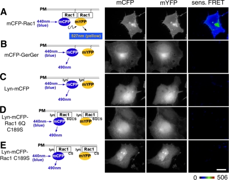
FIGURE 4.
The polybasic region is not sufficient to allow Rac1 self-association of membrane-localized protein. Schematic diagrams on the left represent the pairs of fluorescent proteins used for FRET analysis in COS1 cells. Derivatives used were mCFP-Rac1(WT) and mYFP-Rac1(WT) (A); the mCFP-GerGer and mYFP-GerGer pair having CAAX boxes at the carboxyl termini of the fluorescent proteins, lacking all other Rac1 sequences (24) (B); Lyn-mCFP and Lyn-mYFP, fluorescent proteins localized in the membrane via the NH2-terminal myristoylation site (C); Lyn-mCFP-Rac1-6Q(C189S) and Lyn-mYFP-Rac1-6Q(C189S), localizing hybrid proteins in the membrane via the NH2-terminal myristoylation site (D); and Lyn-mCFP-Rac1(C189S) and Lyn-mYFP-Rac1(C189S), in which CAAX box defective mutants with intact PBRs are forced into the plasma membrane by the Lyn myristoylation signal (E). Constructs encoding these proteins were transfected into COS1 cells, fixed the day after transfection, and subjected to FRET microscopy. Representative images of the CFP and YFP channels, along with the sensitized FRET (sens. FRET) are displayed as color gradient look-up tables, using the displayed scale. White scale bar in E, 10 μm (applies to all panels).

