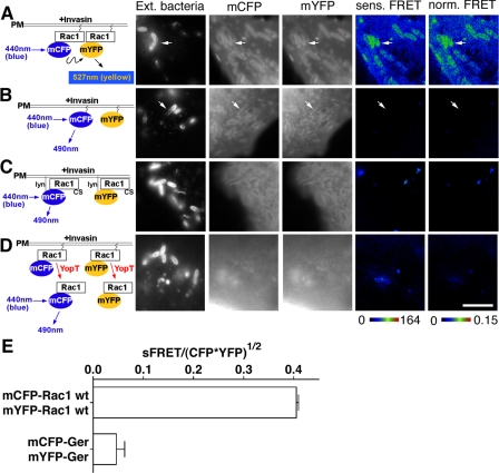
FIGURE 7.
Rac1 self-association is stimulated by Yersinia infection. COS1 cells were transfected with mCFP-Rac1 and mYFP-Rac1 constructs (A and D), mCFP-GerGer and mYFP-GerGer (B), or Lyn-mCFP-Rac1(C189S) and Lyn-mYFP-Rac1(C189S) (C). Transfected cells were challenged the next day with a 30-min incubation of YPIII(p-) (A-C) or YP17/pYopT (D), followed by immunostaining of extracellular bacteria (Ext. bacteria) bound onto host cells. Sensitized and normalized FRET readings (sens. FRET and norm. FRET, respectively) were determined as described (see “Materials and Methods”). The arrows denote nascent phagosomes. E, mean normalized FRET levels at nascent phagosomes were plotted comparing mCFP-Rac1/mYFP-Rac1 and mCFP-GerGer/mYFP-GerGer as described in the legend to Fig. 3I. White scale bar in D, 5 μm (applies to all panels).

