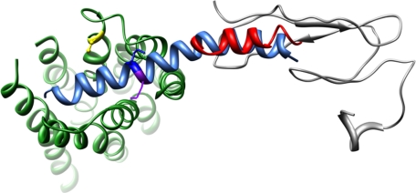
FIGURE 6.
Molecular model of sauvagine binding to CRF1R. The structure of sauvagine, as determined by homology with human urocortin-1, is shown in aqua, with Lys16 and Met17 denoted in dark blue and purple, respectively. The orientation of sauvagine to CRFR1 was guided by the NMR structure of the N terminus of CRFR2 (gray) bound to the C-terminal portion of astressin (red). The 19 residues missing between the CRF2R N terminus as determined by NMR and the beginning of TM1 is denoted by a dashed blue line. Likewise, the EC2 consisting of KLYYDNEKCWFGKRPGVYT is denoted as an orange dashed line, with Cys258 forming a disulfide bond with Cys188 at the extracellular end of TM3 (shown in yellow).

