FIGURE 4.
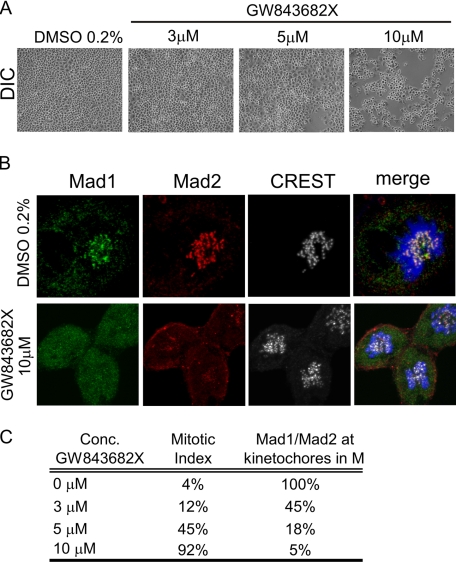
Plk1 inhibitor GW843682X reduced kinetochore association of Mad1 and Mad2. A, light-field images of HeLa cells treated with Plk1 inhibitor GW843682X at various concentrations for 24 h. A dose-dependent increase in cell visually scored as being in mitosis was observed at higher concentrations of GW843682X. B, localization of Mad1 (green) and Mad2 (red) in the presence of GW843682X. CREST (gray) serum was used to visualize centromeres. DAPI is in blue. Sum of z-stack images is shown. A significant reduction in kinetochore association of Mad1 and Mad2 was observed with 10 μm GW843862X. C, an enumeration of the mitotic indices and the frequency of Mad1/2 staining at kinetochores in 0, 3, 5, and 10 μm GW843862X. For kinetochore staining 100 mitotic cells were analyzed in each of the treatment conditions. DIC, differential interference contrast.

