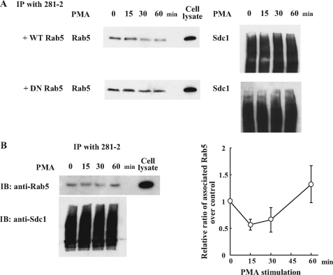
FIGURE 6.
Stimulation of syndecan-1 shedding induces the dissociation of Rab5 from syndecan-1 cytoplasmic domain. A, NMuMG cells transduced with adenovirus harboring either GFP-WT Rab5 or GFP-DN Rab5 were stimulated with PMA (1 μm) for the indicated time periods. Total cell lysate was immunoprecipitated (IP) with 281-2 antibody coupled to Ultralink resins and immunoblotted (IB) with anti-Rab5 or 281-2 antibodies. The band shown is endogenous Rab5 (∼25 kDa), which is readily distinguished from transduced GFP-Rab5 (∼55 kDa). The smear shown is immunoprecipitated syndecan-1. B, NMuMG cells were stimulated with PMA and at the indicated times post-PMA, total cell lysates were prepared, immunoprecipitated with 281-2 antibody-Ultralink beads, and immunoblotted with anti-Rab5 (upper panel) or anti-syndecan-1 antibodies (lower panel with typical proteoglycan smear). The dissociation of Rab5 from the syndecan-1 cytoplasmic domain was quantified by densitometry of the Rab5 band. Data shown are mean ± S.E. from three separate experiments.

