Figure 2.

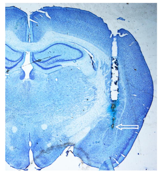
Representative photomicrograph showing the cannula tract and site of injection within the BLA on a cresyl violet stained coronal section. The arrow indicates the most ventral point of the injector cannula tract.

Representative photomicrograph showing the cannula tract and site of injection within the BLA on a cresyl violet stained coronal section. The arrow indicates the most ventral point of the injector cannula tract.