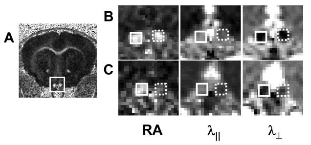
Figure 1.
Mouse optic nerves after one-hour transient retinal ischemia in right eye (reproduced from Sun et al.47 Figure 1 with modification). Optic nerves (rectangle, A) appear hyperintense in a representative diffusion anisotropy, RA, map of a control mouse. Diffusion parameters maps of both control (solid rectangle) and injured (dashed rectangle) optic nerves on 3 (B) and 14 (C) days after retinal ischemia from a representative mouse demonstrating RA, λ∥, and λ⊥ changes after injury. Decreased λ∥ was seen in the injured optic nerve at 3 and 14 days after retinal ischemia, while increased λ⊥ was seen only at 14 days after retinal ischemia.

