
Figure 2.
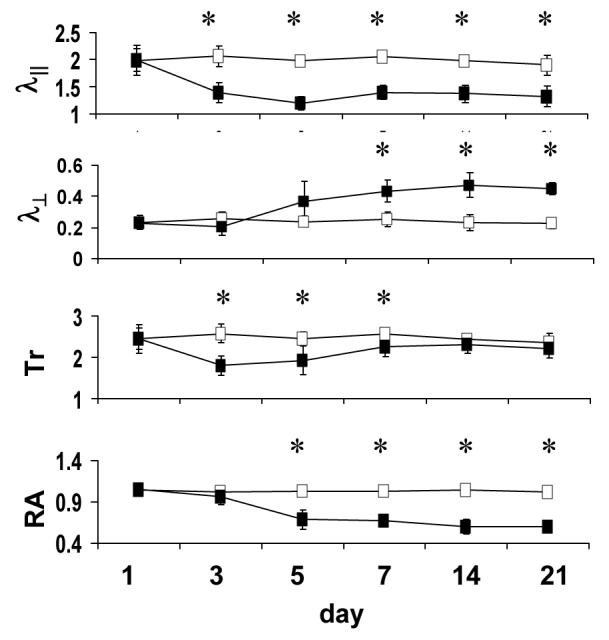
Time course of DTI indices in the optic nerve obtained from mice after transient retinal ischemia (reproduced from Sun et al. 47 Figure 2 with modification). The filled and open squares are measurements obtained from the injured and control optic nerves, respectively. The λ∥ of injured optic nerve decreased significantly at 3 days after reperfusion, remaining at this reduced value throughout the 21-day time course. The λ⊥ of injured optic nerve increased and reached a plateau at 7 days. Decreased Tr was observed at 3 – 7 days as a consequence of reduced λ∥. At 7 – 21 days, decreased λ∥ and increased λ⊥ resulted in the pseudo-normalized Tr. Relative anisotropy decreased at 5 days and remained at the level at 5 – 21 days. Tr, λ∥, and λ⊥ are in units of µm2/ms. RA has no unit. “*” indicates statistically significant difference (p < 0.05) between the control and injured optic nerve.