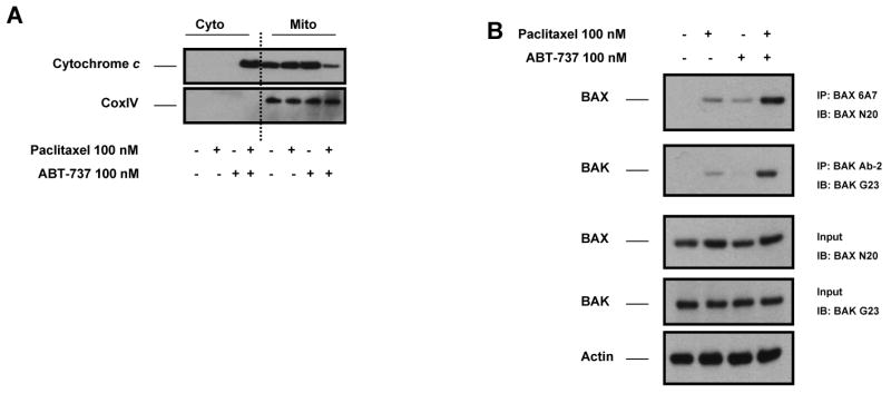
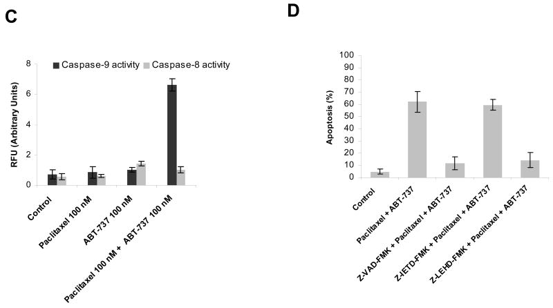
Figure 2.

ABT-737-mediated sensitization of MCF-7 BCL-2 cells to paclitaxel-induced apoptosis via mitochondrial apoptosis pathway. A, MCF-7 BCL-2 cells were treated with paclitaxel (100 nM), ABT-737 (100 nM), or combination of paclitaxel (100 nM) and ABT-737 (100 nM) for 48 h. Cytosolic and mitochondrial fractions from MCF-7 BCL-2 cells were immunoblotted for cytochrome c. CoxIV was probed as a loading control for mitochondrial fractions. B, MCF-7 BCL-2 cells were treated with paclitaxel (100 nM), ABT-737 (100 nM), or the combination of paclitaxel (100 nM) and ABT-737 (100 nM) for 12 h. Activation of BAX and BAK was analyzed by immunoprecipitation with active conformation-specific anti BAX (6A7) and anti-BAK (Ab-2) antibodies followed by immunoblot analysis of BAX and BAK. 5% of the input for immunoprecipitation was also subjected to immunoblot analysis. β-Actin was probed as a loading control. C, MCF-7 BCL-2 cells were treated with paclitaxel (100 nM), ABT-737 (100 nM), or combination of paclitaxel (100 nM) and ABT-737 (100 nM) for 48 h. Activation of caspase-9 and caspase-8 by the combination of paclitaxel and ABT-737 treatment was evaluated by fluorometric caspase activation assays as described in Materials and methods. Data were expressed as relative fluorescence units (RFU). Columns, mean of three independent experiments; bars, SE. D, MCF-7 BCL-2 cells were pretreated with 20 μM pancaspase inhibitor (Z-VAD-FMK), 20 μM caspase-9 inhibitor (Z-LEHD-FMK) and 20 μM caspase-8 inhibitor (Z-IETD-FMK) before treatment with paclitaxel plus ABT-737 for 48 h. The percent apoptotic response was evaluated by Annexin V staining. Columns, mean of three independent experiments; bars, SE.

