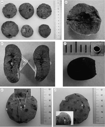
Figure 1.
Type 1 gall fossils. (a) External views (scale in cm). (b) Longitudinal section showing internal airspace, with inner larval chamber missing. The gall's point of attachment is to the left. (c) Two halves of a sectioned compressed gall, showing the larval chamber in the left of the section. (d) A larval chamber, with the adult emergence hole to the right and (inset) in end-on view (scale in mm). (e,f) Two views of the same fossil gall, showing multiple emergence holes (arrowed). In (e), two of the small emergence holes are shown in enlarged view (boxed).

