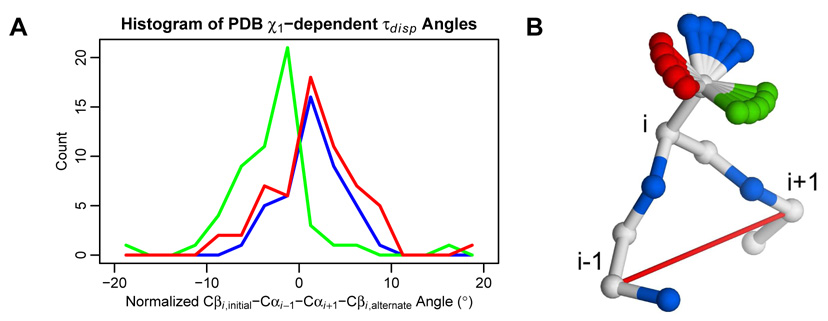
Figure 6.
In high-resolution crystal structures, alternate backbone conformations are correlated with the side chain χ1 angle, with a straightforward structural explanation. A) Out of 126 residues in the backrub set, 68 have χ1 angles in multiple rotameric bins. For those residues, the calculated Cβi,initial–Cαi−1–Cαi+1–Cβi,alternate pseudo-dihedral angles (τdisp) described in Figure 4B were normalized by the average angle (weighted by PDB occupancy). Histograms of those angles are shown using 2.5° bins and colored by χ1 bin: −60° (red), 60° (green), and 180° (blue). B) The clear difference between the −60°/180° and 60° bins has a straightforward structural explanation, where side chains in the 60° bin push the backbone to the left, and the −60°/180° side chains push the backbone to the right. Hypothetical γ atom positions are colored by χ1 bin.

