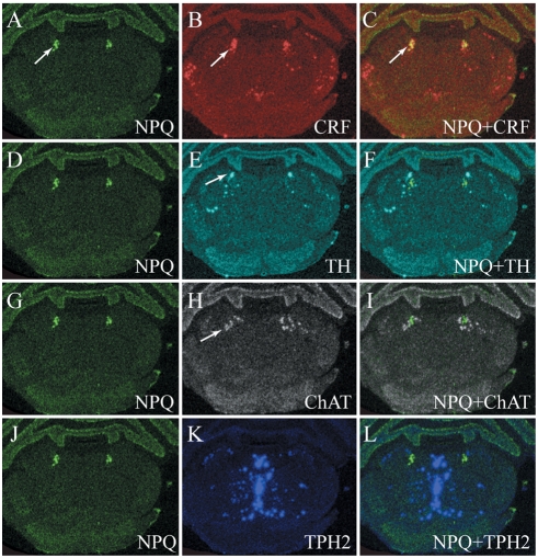
Figure 7. In situ hybridization of preproNPQ mRNA.
Expression of preproNPQ mRNA in the rat brain at the level of Barrington's nucleus and locus coeruleus. In situ hybridizations (ISHs) for preproNeuropeptide Q (NPQ; A, D, G, and J), corticopin-releasing factor (CRF; B), tyrosine hydroxylase (TH; E), choline acetyltransferase (ChAT; H), and tryptophan hydroxylase 2 (TPH2; K) were carried out on adjacent 10 µm-thick sections of the rat brain. ISH autoradiograms were digitized; images were then inverted and pseudocolored according to the following scheme: NPQ – green, CRF – red, TH – cyan, ChAT – white, and TPH2 – blue. To determine whether NPQ signal overlapped with any of the other signals, the sections were aligned and overlaid with each other (C, F, I, L). Arrow in panel A indicates location of NPQ mRNA, while arrow in panel B indicates location of CRF mRNA; note the mixing of red and green (to yield yellow) in panel C (arrow) that suggests co-localization of NPQ and CRF. Arrow in panel E indicates locus coeruleus and its TH-positive neurons. Panel F shows that TH and NPQ signals are spatially very close without overlap. Arrow in panel H indicates the cholinergic laterodorsal tegmental nucleus, while panel I illustrates close spatial relationship between ChAT and NPQ mRNAs. At this level of the neuraxis there is little overlap between TPH2 mRNA (blue signal in panel K, which represents serotonergic neurons) and NPQ (L).

