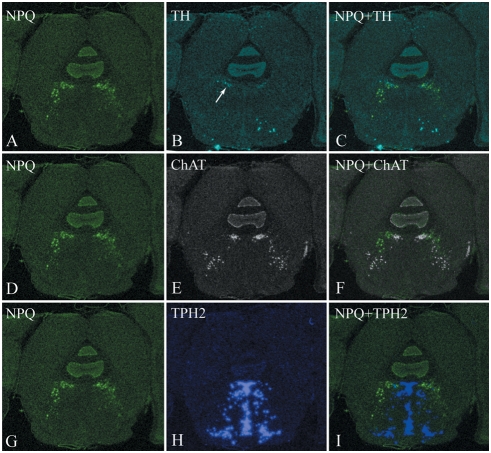
Figure 8. In situ hybridization of preproNPQ mRNA.
Expression of preproNPQ mRNA at the level of the caudal ventrolateral periaqueductal gray (PAG). ISH autoradiograms were digitized and pseudocolored according to the same scheme as in Figure 7. NPQ signal was visible in the ventrolateral quadrant of the PAG as well as within the underlying reticular formation (A, D, G). ISHs for TH (B), ChAT (E) and TPH2 (H) were carried out on adjacent sections. Arrow in panel B indicates location of dopaminergic TH-positive neurons of the ventrolateral PAG that appear to overlap with a subset of NPQ mRNA (C). There is also close spatial relationship between NPQ and ChAT (F) and NPQ and TPH2 (I). Abbreviations are the same as in Figure 7.

