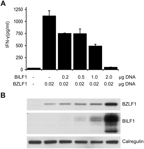
Figure 3. BILF1 inhibits T cell recognition of endogenous EBV antigen in MJS cells.
(A) MJS cells were co-transfected with 0.02 µg p509 plasmid (BZLF1 expression vector) and different amounts (0–2 µg) of pCDNA3-HABILF1-IRES-nlsGFP bulked to a constant amount of DNA with control plasmid. At 24 hr post-transfection, the MJS cells were co-cultured with CD8+ effector ‘RAK’ T cells for a further 18 hrs and the supernatants were tested for the release of IFN-γ as a measure of T cell recognition. All results are expressed as IFN-γ release in pg/ml and error bars indicate standard deviation of triplicate cultures. (B) Total cell lysates were generated from the above transfections, and 2×10⁵ cell equivalents were separated and analyzed by Western Blotting using antibodies specific for BZLF1, HA tag (BILF1), or calregulin as a loading control.

