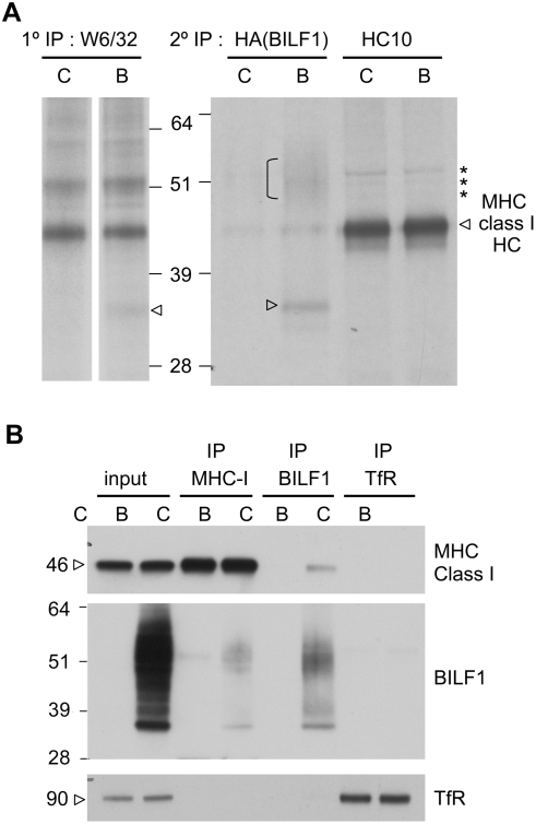
Figure 6. BILF1 is physically associated with the MHC class molecule complex.
(A) 293 cells (107) stably transduced with control (c) or BILF1 (b) retrovirus were metabolically labeled for 15 min and chased for 20 min. After lysis in NP-40 buffer and immunoprecipitation with mAb W6/32, the samples were dissociated by boiling in reducing sample buffer, and were re-precipitated with either 3F10 ( HA tag on BILF1) or HC10 (MHC class I heavy chain). Protein samples were separated by 10% acrylamide SDS/PAGE gel, dried and exposed to autoradiography. The arrowhead and bracket indicate the presence of 33 kD and 45–55 kD BILF1 bands, whilst the asterisks indicate probable ubiquitinated MHC class I species. (B) 293 cells (2×10⁶) stably transduced with control (c) or BILF1(b) retrovirus were treated with concanamycin A (50 nM) for 20 hr prior to lysing the cells with NP40 detergent buffer and immunoprecipitation with antibodies specific for MHC class I (W6/32), HA tagged BILF1 (12CA5), or TfR (H68.4). Cell lysates and immune complexes were separated by SDS/PAGE gel, and analyzed by western blotting using antibodies specific for MHC class I (HC10), HA tagged BILF1 (3F10), and TfR (H68.4). The first two samples on the gel are total cell lysates representing 5% of the input lysate for immunoprecipitations.

